

Which Type Of Procedure Uses An X-ray Beam That Is Controlled By A Computer?
- What is a computed tomography (CT) scan?
- How does CT piece of work?
- When would I get a CT scan?
- What is a CT contrast agent?
- Are there risks?
- What are examples of NIBIB-funded projects using CT?
What is a computed tomography (CT) browse?
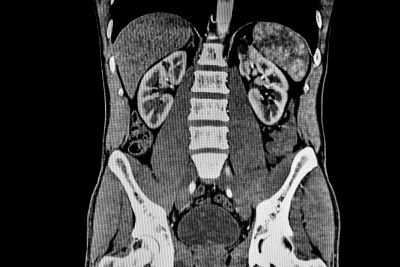
The term "computed tomography," or CT, refers to a computerized x-ray imaging process in which a narrow beam of x-rays is aimed at a patient and quickly rotated around the body, producing signals that are candy past the car's computer to generate cantankerous-sectional images, or "slices." These slices are called tomographic images and tin can give a clinician more detailed information than conventional x-rays. In one case a number of successive slices are nerveless by the auto'due south figurer, they tin exist digitally "stacked" together to form a three-dimensional (3D) image of the patient that allows for easier identification of basic structures also as possible tumors or abnormalities.
How does CT work?

Dissimilar a conventional x-ray—which uses a stock-still x-ray tube—a CT scanner uses a motorized x-ray source that rotates around the round opening of a donut-shaped structure called a gantry. During a CT browse, the patient lies on a bed that slowly moves through the gantry while the ten-ray tube rotates around the patient, shooting narrow beams of 10-rays through the body. Instead of picture show, CT scanners apply special digital ten-ray detectors, which are located directly opposite the ten-ray source. As the ten-rays go out the patient, they are picked up by the detectors and transmitted to a figurer.
Each time the 10-ray source completes one total rotation, the CT calculator uses sophisticated mathematical techniques to construct a 2-dimensional epitome slice of the patient. The thickness of the tissue represented in each image slice can vary depending on the CT machine used, but usually ranges from 1-10 millimeters. When a full slice is completed, the image is stored and the motorized bed is moved forward incrementally into the gantry. The x-ray scanning process is then repeated to produce another paradigm piece. This procedure continues until the desired number of slices is nerveless.
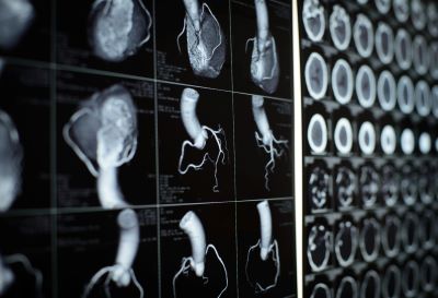
Prototype slices can either be displayed individually or stacked together by the computer to generate a 3D paradigm of the patient that shows the skeleton, organs, and tissues as well every bit whatever abnormalities the dr. is trying to identify. This method has many advantages including the ability to rotate the 3D prototype in infinite or to view slices in succession, making it easier to discover the exact place where a problem may be located.
When would I get a CT browse?
CT scans tin be used to identify disease or injury within various regions of the body. For example, CT has go a useful screening tool for detecting possible tumors or lesions within the abdomen. A CT browse of the heart may be ordered when various types of heart affliction or abnormalities are suspected. CT can as well exist used to image the head in order to locate injuries, tumors, clots leading to stroke, hemorrhage, and other conditions. Information technology tin image the lungs in social club to reveal the presence of tumors, pulmonary embolisms (blood clots), excess fluid, and other conditions such as emphysema or pneumonia. A CT scan is peculiarly useful when imaging complex bone fractures, severely eroded joints, or bone tumors since it usually produces more detail than would exist possible with a conventional x-ray.
What is a CT contrast agent?
As with all x-rays, dense structures inside the body—such equally bone—are hands imaged, whereas soft tissues vary in their power to end x-rays and therefore may exist faint or difficult to run across. For this reason, contrast agents have been developed that are highly visible in an x-ray or CT browse and are safe to employ in patients. Dissimilarity agents contain substances that can stop x-rays and are therefore more visible on an 10-ray image. For instance, to examine the circulatory system, an intravenous (Iv) contrast agent based on iodine is injected into the bloodstream to aid illuminate blood vessels. This type of test is used to await for possible obstructions in blood vessels, including those in the heart. Oral contrast agents, such as barium-based compounds, are used for imaging the digestive system, including the esophagus, stomach, and gastrointestinal (GI) tract.
Are there risks?
CT scans tin can diagnose maybe life-threatening weather such as hemorrhage, blood clots, or cancer. An early diagnosis of these conditions could potentially be lifesaving. However, CT scans utilise x-rays, and all x-rays produce ionizing radiation. Ionizing radiation has the potential to cause biological furnishings in living tissue. This is a run a risk that increases with the number of exposures added up over the life of an private. Nonetheless, the chance of developing cancer from x-ray radiations exposure is more often than not small.
A CT scan in a pregnant woman poses no known risks to the infant if the area of the body being imaged isn't the abdomen or pelvis. In full general, if imaging of the belly and pelvis is needed, doctors prefer to use exams that practice not use radiation, such as magnetic resonance imaging (MRI) or ultrasound. Nonetheless, if neither of those can provide the answers needed, or at that place is an emergency or other time constraint, CT may be an adequate alternative imaging choice.
In some patients, contrast agents may cause allergic reactions, or in rare cases, temporary kidney failure. IV contrast agents should not be administered to patients with aberrant kidney part since they may induce a farther reduction of kidney function, which may sometimes become permanent.
Because children are more sensitive to ionizing radiation and have a longer life expectancy, they have a higher relative risk for developing cancer from such radiation compared with adults. Parents may want to ask the technologist or doctor if their machine settings accept been adjusted for children.
What are examples of NIBIB-funded projects using CT?
Imaging for acute ischemic stroke: Stroke, which can have lasting neurological injuries, is also a leading crusade of decease worldwide. To mitigate impairment to the brain, patients may receive endovascular treatment, where the clot blocking the claret supply is either removed or dissolved. Notwithstanding, identifying patients who will benefit from endovascular therapy, such equally those with just a modest volume of irreversibly injured brain tissue, remains challenging, and fourth dimension is a critically important cistron for a successful clinical outcome.

NIBIB-funded researchers accept adult an epitome reconstruction technique to more efficiently triage patients who present with symptoms of a stroke. This CT-based method can exist used to rule out the presence of a hemorrhage; to find the site of the blood clot; and to identify the extent of damaged brain tissue. Such a technique could significantly shorten the time from the diagnosis of a stroke to the first of endovascular therapy, and could also guide the endovascular treatment. Following evaluation in animal models, researchers program to validate this CT imaging technique in human being studies.
Accounting for metal implants in CT imaging: Metallic objects, such as implants and prostheses, can innovate 'artifacts' that may announced as streaks or shadows on a CT scan. These artifacts can obscure anatomical structures or impact calculations necessary for planning radiation therapy. While techniques be to reduce such artifacts, they practice not fully mitigate the artifacts and may fifty-fifty introduce new ones. In this projection, NIBIB-funded researchers have developed an algorithm to reduce metallic artifacts in CT imaging, without requiring knowledge of the implant material. The researchers plan to optimize their algorithm and and then evaluate their technique every bit a potential method to better radiations therapy planning for prostate cancer amidst those with hip protheses.
Leveraging CT images to guide treatments for COVID-19 and beyond: Artificial intelligence is increasingly being used with medical imaging, such as CT, to assist meliorate diagnoses and guide treatment decisions. By using medical images and patient outcomes, clinicians tin can "railroad train" machine learning-based technologies to recognize patterns and predict responses. During the COVID-19 pandemic, NIBIB created a collaborative imaging initiative called the Medical Imaging and Data Resource Center (MIDRC). This initiative collected and analyzed thousands of CT images from patients with COVID-nineteen for the development of artificial intelligence and auto learning tools to guide the treatment and monitoring of the affliction. These datasets contribute to the development of algorithms for detection, prognosis, and optimization of therapy in astute COVID-xix patients and have the potential to contribute to the understanding of Mail service-Acute Sequelae of SARS-CoV-2 infection (PASC, otherwise known equally "Long COVID"). Further, this initiative paves the way for new tools that leverage imaging for other medical atmospheric condition, such every bit cancer, liver disease, or other infectious diseases, amid others.
For more information about CT, watch our video here.
Updated June 2022

Which Type Of Procedure Uses An X-ray Beam That Is Controlled By A Computer?,
Source: https://www.nibib.nih.gov/science-education/science-topics/computed-tomography-ct
Posted by: backmanwhourpel.blogspot.com
0 Response to "Which Type Of Procedure Uses An X-ray Beam That Is Controlled By A Computer?"
Post a Comment